Canine Cruciate Ligament (CCL / ACL) Repair Surgery TPLO & Extracapsular Repairs
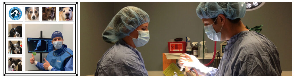
Dr. Fritz Trybus at Cary Grove Animal Hospital has been performing cruciate ligament repair surgery (CCL/ACL) in dogs since 2007. He is trained in both the extracapsular/lateral suture procedure and the Tibial Plateau Leveling Osteotomy (TPLO). We are proud to be one of the only animal hospitals in the McHenry County, Illinois area offering the Tibial Plateau Leveling Osteotomy (TPLO) surgery for dog cruciate (CCL / ACL) ligament tear surgery. We are able to perform surgery in dogs up to 150 pounds.
To set up a surgery consultation for your dog with Dr. Fritz please call us at 847-462-8387 or e-mail [email protected]. We are able to schedule most standard dog cruciate surgeries within 2 weeks for extracapsular repair or TPLO surgery. Please bring your dog fasted (no food for 12 hours, a small amount of water is fine) to your first visit as mild sedation may be given prior to radiographs (x-rays) that we may need to take.
We also utilize a board-certified traveling surgeon that works out of our hospital for more complicated orthopedic surgeries including fracture repair. These fees are quoted on an individual basis.
What is the estimated cost for an ACL surgery at Cary Grove Animal Hospital?
Setting up an examination and consultation is the best way for us to provide you with an accurate estimate of costs. All patients are required to have a pre-surgical examination, blood test and x-rays prior to surgery (not included in surgical cost).
All surgeries include balanced anesthesia, pain management, medications to go home with (antibiotics, pain medications), and follow up consultations and recheck x-rays. Package Surgery Costs are:
* Extracapsular Surgery Cost Estimate: $3,200
* TPLO Surgery Cost Estimate: $4,800 (giant breed dogs over 90 lbs may have additional charges depending on implants needed)
Canine Cruciate Ligament (CCL / ACL) Tear Information
Rear leg lameness or limping in the dog is a common problem and concern for all types of dog owners. There are many orthopedic and soft tissue injuries and conditions that can occur in the rear limbs of canines. The knee or stifle joint is a major source for these problems, especially rupture of the Cranial or Anterior Cruciate Ligament. This article will focus on an introduction to Cruciate Ligament ruptures and their cause, how a diagnosis is made, and the current treatment options available.
Cranial Cruciate Ligament (CCL or ACL) rupture is the most common knee condition that we see in dogs today. It is diagnosed in all sizes of dog breeds from Great Danes to Chihuahuas.
The ACL is an important structure in providing stability to the rear limb and limiting internal rotation. While many times the cruciate rupture appears to be associated with a traumatic event or injury, we now know that the majority of ACL ruptures have many contributing factors.
This problem is often more accurately referred to as Cruciate Disease. The conditions within the dog’s joint, conformation and angles of the joint and genetic factors all play a role. In addition, obesity and lack of conditioning will contribute to rupturing of the ACL.
Any time a dog is non-weight bearing or partial weight bearing of a rear limb, an ACL rupture is high on the list of possible causes. ACL ruptures may occur both acutely or more chronically. In some cases the limping is more subtle and waxes and wanes over a period of weeks or months.
The ACL may either rupture partially or fully. This creates a great deal of inflammation within the joint causing pain and discomfort. Both full and partial ruptures are best treated with surgery. While many dogs do not whine or cry, if they are limping on a leg we know there is discomfort. Cases with full ruptures have a large amount of instability within the joint. In about 50% of these cases the dog may also have a rupture of the meniscus, another ligament within the knee in close relation to the ACL. Meniscal tears must be addressed at the time of surgery by removing the torn or damaged portion. Many of the dogs with Cruciate Disease in one leg will at some point have the other leg affected. This may be at the same time as the first leg, or months to years later.
Diagnosis of a cruciate rupture involves a few steps. When presented with lameness of a rear leg, the veterinarian will palpate the knee carefully. Evaluating the knee includes feeling for swelling or effusion within the joint. A natural indentation should be felt next to the patellar tendon in the stifle. Many times with effusion of the joint, this natural divot or indentation is no longer present due to joint fluid swelling and bulging.
The veterinarian will also test for cranial drawer sign and tibial thrust. This may need to be done under sedation depending on the dog. It involves being able to move the tibia cranially or forward in relation to the femur. If positive we know the cruciate ligament is ruptured. However, not all cases of cruciate disease will elicit a positive cranial drawer test. Cases of partial ruptures, which may cause the dog just as much discomfort as a full tear, may not show any instability on the cranial drawer test.
Observing the dog walk, trot, and sit will also help make a diagnosis. The “sit” test is one where the dog with a ruptured ACL will not sit squarely anymore. They instead have a more puppy-like sit or a “sloppy” sit. They will kick one or both rear legs out, extending them forward or off to the side. They will not sit squarely with a normal bend to the knees.
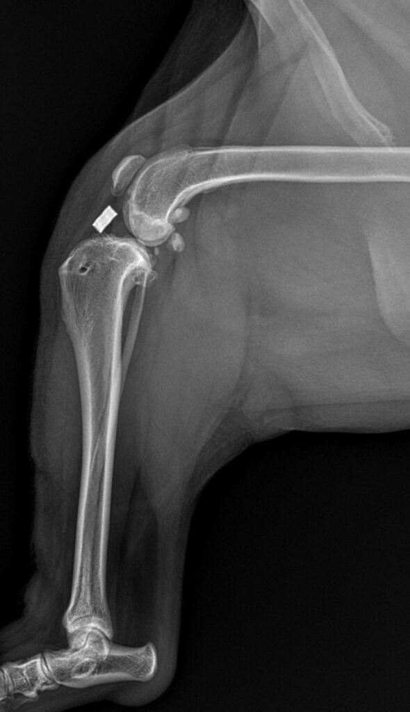
When dealing with a dog that you suspect has an ACL tear, seeking a veterinarian to evaluate them is important. Physical exam findings are correlated with radiographic findings. While ligaments do not show up on plain radiographs (x-rays), there are specific changes that are seen radiographically that can help confirm a diagnosis of an ACL rupture. It is also important to help rule out other conditions with the radiographs, which may have similar clinical presentations. These other conditions include hip dysplasia, muscular strains, and bone cancer. However, in some cases there may be more than one orthopedic condition at play at the same time. It is not uncommon to reveal hip dysplasia with radiographs, but also find an ACL rupture! Most often, the ACL rupture is what is creating the majority of lameness if the problem presented more acutely.
With ACL ruptures being so common there are many different surgical procedures that are available for their repair. Some question the need for surgery at all and may advocate just rest and rehabilitation. Research has proven that dogs that undergo ACL surgery have a far superior outcome than dogs that do not have surgery. Without surgery joint stabilization may never occur, damaged ligaments will create discomfort if not cleaned out of the joint, and premature arthritis will set in. Additionally, the dog will stress out their “normal” leg much more possibly leading to another ACL rupture and will experience muscle atrophy and pain in other joints. Braces or bandages are not effective in dogs for healing ACL ruptures or stabilizing the knee joint.
The first part of all knee surgery is to enter the joint and evaluate all structures. This is done with an arthrotomy or incision into the joint. Damaged ligaments are removed and the joint is cleaned of debris. Meniscal ligaments are also evaluated at this time.
There are two major groups of surgeries: extracapsular stabilization (or often called “lateral suture” technique) and geometry modifying procedures (the gold standard being a TPLO).
Extracapsular Repair
Extracapsular repairs are the most traditional way to stabilize the knee joint after ACL rupture. These surgeries involve placing an implant outside of the joint to act as the ACL would to eliminate instability. It involves a suture that is placed around the lateral femoral-fabellar ligament through a hole drilled in the tibial crest area. This procedure has many variations. Some veterinarians use fishing-line like material, others a braided polyester suture. Some surgeons tie knots to secure the suture, others may use a crimping system to eliminate a large knot placement. There are also variations on the location of placement of the bone tunnel through the tibia, depending on surgeon preference.
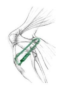
Tibial Plateau Leveling Osteotomy (TPLO)
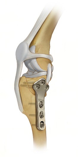
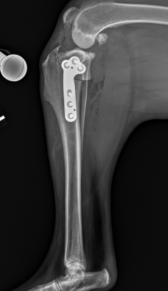
Geometry modifying procedures are most commonly done for dogs weighing over 40 pounds who may not achieve adequate stabilization with the more basic extracapsular repair technique. The most common surgeries within this category are the Tibial Plateau Leveling Osteotomy (TPLO) and Tibial Tuberosity Advancement (TTA). The TPLO is the gold standard as it provides the best short and long term results of all Cruciate Ligament Repair surgeries. A TPLO involves cutting the tibia to change the slope of the joint, thus changing the mechanics within the knee to provide stabilization. Bone plates are placed on the newly cut bone to secure the angles and allow the bone to heal. These bone plates are generally left in place for the life of the dog.
The TPLO surgery offers excellent long term stabilization and functionality of the repaired limb and is the gold standard procedure for most large and/or active dogs. A drawback of the TPLO is cost, as these are more expensive than the extracapsular repair surgery.
Following surgery, about 8-10 weeks of rest/reduced activity is required and an additional 4-6 week period to slowly increase activity of the patient. During the recovery phase it is very important to keep the patients on a leash at all times and avoid running, jumping, or access to stairs. Release instructions will be given to owners after the surgery outlining what the dog is allowed to do and not do. Some dogs may require structured rehabilitation including underwater treadmill exercise if they do not progress adequately. Most dogs however only require at home care during the recovery phase with controlled exercise. We also give the option to incorporate therapy LASER treatments for the first 2-3 weeks after surgery with patients at Cary Grove Animal Hospital to help with the healing process.
As outlined, there are many aspects of cruciate ligament ruptures in dogs. The causes of the rupture are often multi factorial and there are many methods that are used to reach an accurate diagnosis. Multiple factors go into deciding which surgery is best for the individual dog and owner. Regardless of the method of repair chosen, good communication between the owner and veterinarian and diligent at home post-operative care is of upmost importance to achieve a favorable recovery.
Essential Preventative Care Services:
Protection against rabies, distemper, parvo, and other diseases.
Year-round protection against heartworm, fleas, ticks, and intestinal parasites
Evaluation of oral health with vet recommendations on dental care.
Dietary recommendations for optimal health.
Blood and urine tests to detect hidden health issues.
Permanent identification if your pet gets lost.
Solutions for common behavior problems.
When Does Your Pet Need A Wellness Exam?
- Puppies And Kittens: Multiple visits during first year for vaccines and development checks.
- Adult Dogs And Cats (1 — 7 Years): Annual comprehensive exams.
- Senior Pets (7+ Years): Twice-yearly exams with additional screening tests.
- After Adoption: Within first week of bringing home a new pet.
- Before Boarding Or Grooming: To ensure your pet is healthy and current on vaccines.
Tailored Care For Every Life Stage:
- Puppy & Kitten
- Adult Pet
- Senior Pet
- Series of vaccinations and boosters
- Deworming treatments
- Growth and development monitoring
- Nutritional guidance for proper development
- Spay/neuter planning
- Training and socialization advice
- Breed specific advice
- Annual physical examinations
- Vaccination boosters as needed
- Dental health assessment
- Weight management
- Parasite prevention
- Semi-annual examinations
- Senior blood profiles
- Arthritis and mobility assessments
- Age-appropriate nutritional counseling
- Disease screening for common senior conditions
Book Your Pet's Wellness Exam Today
Taking care of your pet’s health starts with a thorough veterinary exam. To schedule an appointment for your pet, call us at 847-462-8387.
Bringing You Peace of Mind
We are committed to sustaining a healthy lifestyle during all facets of your pet’s milestones

Unparalleled Experience
Compassionate quality care in Cary, Illinois and surrounding cities CHANGE!!!!!
Comprehensive Care
We offer a variety of services for pets of all ages, including dentistry, surgery, and wellness care.
Pet Life Commitment
We are here to provide your pets with excellent care, not only during emergencies but for all of their wellness needs.